
High-Content Screening (HCS)
High-Content Screening from Target ID to Candidate Selection
HCS is a high-throughput whole-cell imaging platform that enables compounds screening in complex cellular systems to identify promising leads or reveal new MoA. This advanced approach can help identify potential first-in-class drugs for currently intractable conditions. As you progress your candidate through lead optimization, you can use high-content imaging to monitor compounds responses in complex cellular systems to assess their potential for translational success in vivo.
WuXi Biology and Testing’s high-content imaging (HCI) and high-content screening (HCS) platform brings together cell biology and in vitro toxicology expertise with laboratory automation, compound management, and image analysis capabilities. This integrated platform enables high-throughput imaging and multiparametric analysis across complex cellular systems to support diverse screening applications.
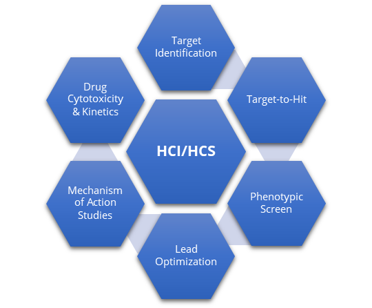
High Content Imaging as an Early Discovery Tool
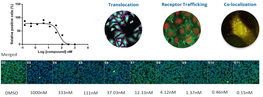
HCS is a high-throughput whole-cell imaging format that enables the screening of thousands of compounds in complex cellular systems to identify new drug targets or new lead compounds. Some examples are below.
Assay Applications
High-content imaging supports a broad range of cellular and phenotypic assays, from target and pathway characterization to compound efficacy and in vitro safety assessment.
- Cell Painting
- Immunohistochemistry
- RNAi
- Reporter gene assays
- Protein-protein interactions
- Cell Proliferation and cytotoxicity
- Cell cycle
- Autophagy
- Protein degradation
- DNA damage
- Morphological changes
- Protein expression
- Protein localization
- Post-translation modifications
- Developmental toxicity
- Genotoxicity (e.g., micronucleus formation)
- Neurotoxicity (e.g., neurite outgrowth)
- Hepatotoxicity (e.g., steatosis, phospholipidosis)
- Cardiotoxicity (e.g., mitochondrial oxidative stress, mitochondrial membrane potential)
- Nephrotoxicity
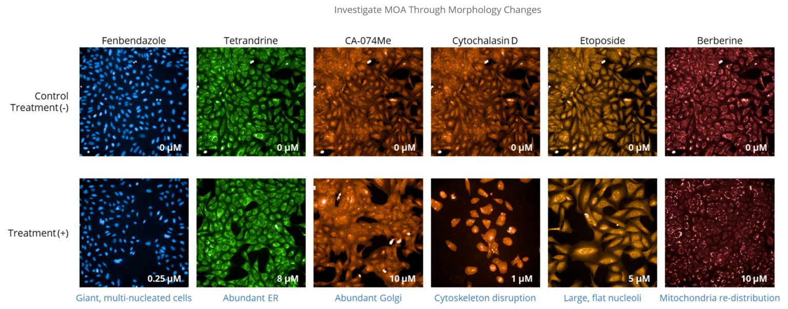
Case Study: Phenotypic Screen Using Cell Painting
Cell painting was performed in U-2 OS osteosarcoma cells using high-content, high-throughput imaging to capture a broad range of morphological features in response to diverse perturbations. This multiparametric approach enabled unbiased phenotypic profiling and could be adapted to specific screening conditions to support compound characterization, as well as the evaluation of potential toxicity.

Explore Solutions for Your Program
Explore solutions by development phase, drug modality, therapeutic area, or integrated discovery needs.
You May Also Be Interested In



Get a Quote
Ready to plan your next study? Share your program needs with our team for a tailored estimate.